A mouse study reveals something counterintuitive: losing just a small segment of myelin—the protective coating around nerve fibers—can completely derail how the brain processes information, even when the rest of the signal gets through.
Nerve cells rely on myelin like fiber-optic cables rely on insulation. It speeds up electrical signals so the brain can think and sense in real time. Scientists knew this much. What they didn't know was what happens when that insulation breaks down in just one critical spot—and it turns out, the consequences are surprisingly severe.
Researchers at Maarten Kole's lab ran experiments in mice, focusing on the communication pathway between the brain's outer layer (the cerebral cortex) and the thalamus, a relay station buried deep inside. This back-and-forth conversation—called the corticothalamic loop—is how your brain processes touch, sight, and sound. In humans, the same loops underpin everything from sensory perception to memory and attention.
We're a new kind of news feed.
Regular news is designed to drain you. We're a non-profit built to restore you. Every story we publish is scored for impact, progress, and hope.
Start Your News DetoxThe missing first signal
When the team deliberately degraded myelin in a targeted way, they expected the entire nerve fiber to lose its insulation. Instead, only the sections nearest the cell body were affected. This mimics what happens in Multiple Sclerosis, particularly in gray matter lesions, where cognitive symptoms tend to be most severe.
Here's where it gets interesting. The mice could still send signals to the thalamus—but the signals had lost their "first wave." Kole uses a barcode analogy: imagine a supermarket scanner that needs to read every stripe to identify a product. Skip the first stripe and the whole scan fails. The signal gets through, but the brain can't decode it.
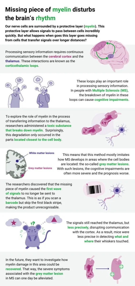
When a mouse's whiskers touch an object, the cortex normally amplifies the thalamus signal to help the brain pinpoint exactly what and where it's touching. With the myelin gap, that amplification still happened—but less accurately. The mouse could feel something, but couldn't quite identify what it was or when it happened. The communication loop between the two brain areas broke down.
Why this matters
This finding explains something that has puzzled neurologists for years: why gray matter lesions in MS cause such severe cognitive problems—disorientation, driving difficulties, trouble recalling familiar names—even when the overall signal seems to get through. The brain isn't just receiving slower messages; it's receiving messages it can't interpret. The code changes, and the whole system loses track.
Kole's team is now investigating whether myelin damage in this critical area can be repaired. If they can restore those first signals, it might one day ease some of the most debilitating symptoms of MS. The work appears in Nature Communications and opens a new angle on how the brain's wiring works—and what happens when even tiny pieces of it come loose.