

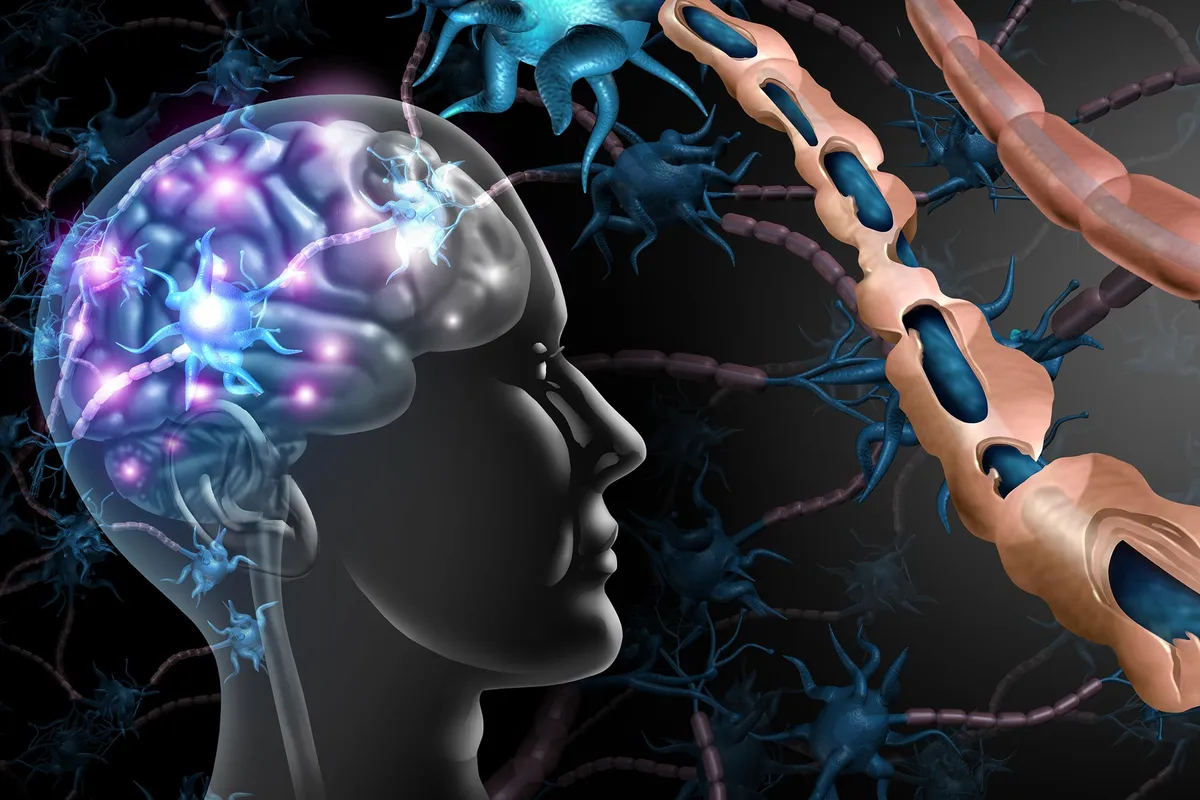
For years, doctors trying to understand Multiple Sclerosis (MS) were essentially flying blind when it came to the brain's gray matter. They could see the obvious damage in the white matter on an MRI, but a whole universe of crucial lesions — linked to disability and cognitive decline — remained stubbornly invisible.
Now, an international team, spearheaded by the University at Buffalo, has taught AI to find what human eyes (and traditional software) couldn't. It's like turning on a blacklight in a dimly lit room, revealing all the things you really didn't want to know were there, but absolutely needed to see.
Your Old Scans Just Got a New Life
This isn't about fancy new MRI machines; it's about making existing ones work harder. The AI method takes multiple standard MRI images and cross-references them, sniffing out disease signals that were previously too subtle to detect. The breakthrough, published in Communications Medicine, means a more complete picture of MS progression, particularly those insidious thinking problems and physical disabilities.
We're a new kind of news feed.
Regular news is designed to drain you. We're a non-profit built to restore you. Every story we publish is scored for impact, progress, and hope.
Start Your News DetoxDr. Robert Zivadinov, one of the study's senior authors, put it plainly: this discovery has "big implications." Because apparently that's where we are now — AI is looking at your old scans and finding things your neurologist couldn't. Which, if you think about it, is both impressive and slightly terrifying.
Cortical lesions, those tiny problem areas in the brain's outer layer, have long been suspected culprits in MS. But without a reliable way to see them, they were useless for diagnosis or monitoring. Dr. Michael G. Dwyer, the lead author, expressed the frustration perfectly, noting that a "lot of ongoing damage in MS wasn’t visible with traditional MRI." That is, until now.
The AI's Secret Sauce
The researchers built on earlier work from Dutch coauthors, combining various image processing methods, including a new one called MMCLE (multimodal cortical lesion enhancement). They then unleashed these methods on MRI scans from the ORATORIO clinical trial, which involved over 700 MS patients.
What they found was a hidden layer of damage. While standard scans showed the usual white matter lesions, the AI-guided processing revealed an average of 15 to 20 cortical lesions per patient. That's over 11,000 previously unseen lesions across the entire dataset. Let that satisfyingly large number sink in.
Dr. Dwyer explained that generative AI's power lies in its ability to spot the tiny differences between scans that indicate unhealthy tissue. It's like a digital detective noticing the faint smudge that reveals the whole story. This means doctors can now pull incredibly useful data from existing scans, giving them a much clearer battlefield map for MS.
This international effort, involving scientists and doctors from multiple universities and companies (including Genentech), is set to significantly impact how clinical trial data — past, present, and future — will be reviewed. Your brain scans just got a second, much more thorough, opinion.