Researchers at Caltech and USC have developed a way to see inside the human body that does something existing scans can't: show both the structure of soft tissue and how blood is moving through it, all at once, in full color.
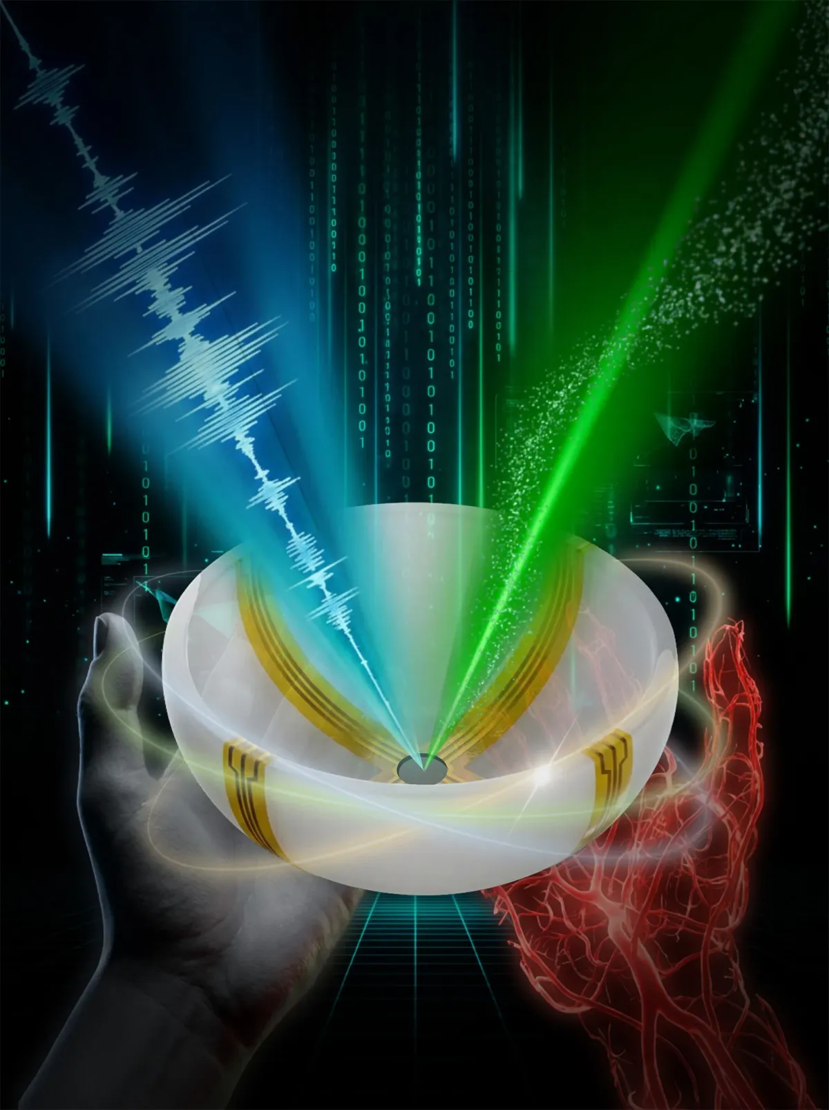
The technique is called RUS-PAT, and it works by combining two imaging methods that have existed separately for years. One uses ultrasound waves to map tissue structure. The other, photoacoustic imaging, uses laser pulses to make molecules vibrate and produce sound waves that reveal blood vessels and oxygen levels. By merging these two approaches, the researchers created something more informative than either alone.
What makes this practical is the engineering. Earlier attempts to combine these methods required so many sensors and so much complexity that a clinical version seemed unrealistic. The Caltech team found a simpler path: use a single wide-field ultrasound transducer to broadcast waves into the tissue, then use a small number of arc-shaped detectors rotated around the area to capture the returning signals. It's elegant enough to actually work in a hospital setting.
We're a new kind of news feed.
Regular news is designed to drain you. We're a non-profit built to restore you. Every story we publish is scored for impact, progress, and hope.
Start Your News DetoxIn early tests on different parts of the human body, the system produced three-dimensional images in less than a minute, scanning to a depth of about 4 centimeters. The researchers see clear applications. For breast cancer, doctors could see a tumor's exact location and shape alongside its blood supply patterns — information that matters for treatment planning. For patients with diabetic nerve damage, a single scan could show both nerve structure and oxygen delivery. In brain imaging, it could reveal structural detail while tracking blood flow, something neuroscientists have wanted for years.
The depth limitation isn't permanent. Light can be delivered through an endoscope, potentially opening access to deeper tissues. The speed — under a minute per scan — also matters practically. Faster scans mean patients move less, images stay clearer, and doctors can repeat them more often to track changes over time.
Lihong Wang, who developed photoacoustic imaging two decades ago, led the work. His insight was deceptively simple: instead of trying to add ultrasound generation to the existing system, what if you just mimicked light excitation using ultrasound itself. That one question led to a design that's both more capable and more feasible than what came before.
The next phase is moving from demonstration to clinical use. The National Institutes of Health has already funded the work, signaling confidence in the approach. What happens next depends on whether hospitals can integrate this into their imaging workflows — and early signs suggest they can.