Researchers at the University of Surrey have built an AI system that does something doctors have never been able to do before: show a patient what their knee might look like a year from now.
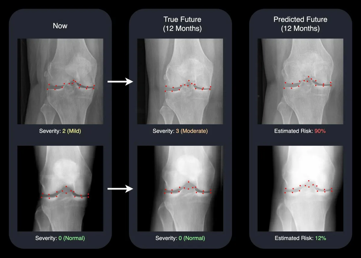
The technology generates a realistic X-ray image of a patient's future joint, paired with a score estimating how likely the disease is to progress. It's the difference between a doctor saying "your arthritis might get worse" and actually showing you what that means on an image side by side with today's scan.
Why This Matters for 500 Million People
Osteoarthritis affects more than 500 million people globally and is the leading cause of disability among older adults. For decades, doctors have relied on numbers and clinical scores to explain disease progression — abstract data that doesn't always stick. A patient might nod along during an appointment, then ignore lifestyle changes because the risk feels theoretical. Seeing a predicted future knee changes that calculation.
We're a new kind of news feed.
Regular news is designed to drain you. We're a non-profit built to restore you. Every story we publish is scored for impact, progress, and hope.
Start Your News DetoxDavid Butler, the study's lead author, puts it plainly: "Seeing the two X-rays side by side — one from today and one for next year — is a powerful motivator. It helps doctors act sooner and gives patients a clearer picture of why sticking to their treatment plan or making lifestyle changes really matters."
The system was trained on nearly 50,000 knee X-rays from almost 5,000 patients, one of the largest osteoarthritis datasets ever used for this purpose. What makes it clinically viable isn't just accuracy — it's speed and simplicity. The AI predicts disease progression more accurately than existing tools while operating around nine times faster and in a more compact form, meaning it could actually fit into everyday hospital workflows rather than sitting in a research lab.
Building Trust Through Transparency
The system uses a type of generative AI called a diffusion model to create the projected X-ray and marks 16 key points within the joint. By showing exactly which areas the AI is tracking for change, clinicians can see the reasoning behind the prediction. That transparency matters. Medical staff are skeptical of black-box AI for good reason — they need to understand what they're looking at before they use it to guide patient care.
Gustavo Carneiro, Professor of AI and Machine Learning at Surrey, notes that earlier systems "were often slow, opaque, and limited to numbers rather than clear images. Our approach takes a big step forward by generating realistic future X-rays quickly and by pinpointing the areas of the joint most likely to change."
The research was presented at the International Conference on Medical Image Computing and Computer Assisted Intervention (MICCAI 2025). The team is now looking for partners to bring the technology into real clinical settings — the next critical step between promising research and actual patient benefit.
The implications extend beyond knees. The same approach could eventually predict lung damage in smokers or track heart disease progression, giving patients visual insight that supports earlier intervention across multiple chronic conditions.