Imagine trying to build a 3D model of something in your head, but all you've got are flat, 2D slices. That's essentially what medical professionals do every time they read an ultrasound. It's a skill that takes years to master, and frankly, sounds a bit like a superpower.
But what if you actually had X-ray vision? Researchers at MIT have cooked up an augmented reality system that lets users see a precise 3D digital model of what they're scanning, making those tricky tissues about a thousand times easier to identify. It's like strapping on a VR headset and suddenly having Superman's eyes, but for your internal organs.
Goodbye, Guesswork
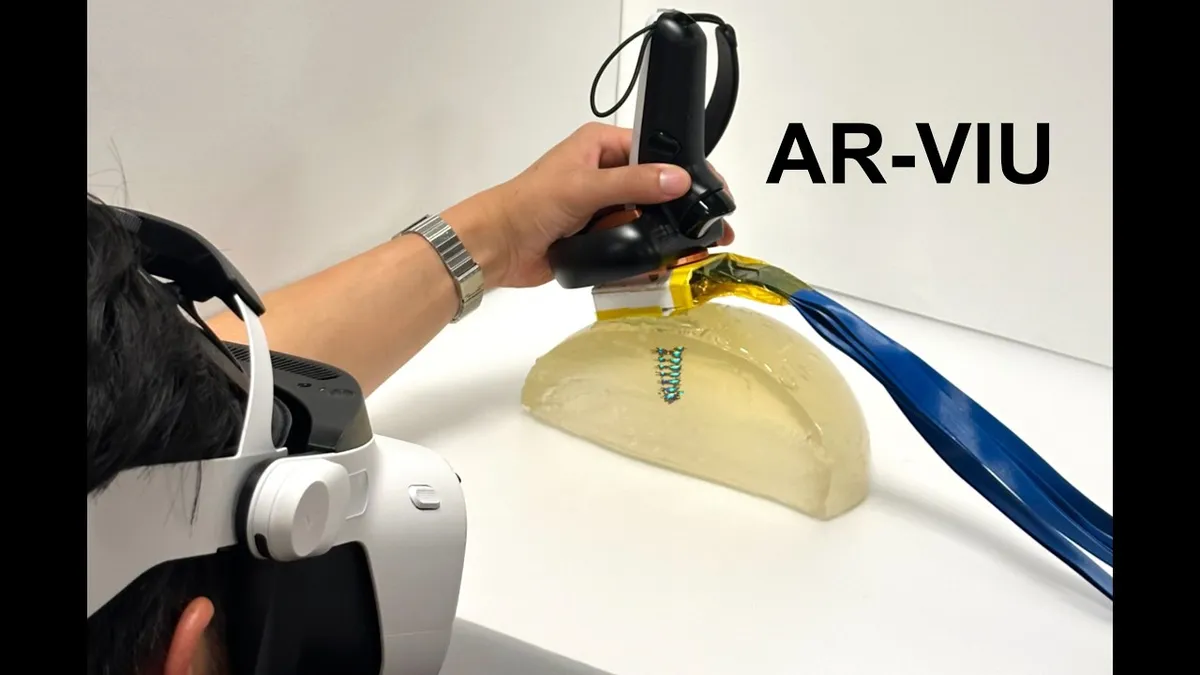
This new tech, dubbed AR-VIU (augmented real-time volumetric imaging in ultrasound), isn't just a party trick. It could dramatically speed up training for ultrasound technicians, help doctors guide biopsy needles with pinpoint accuracy, and generally reduce the stress of trying to diagnose someone based on what looks like a blurry photo of a cloud.
We're a new kind of news feed.
Regular news is designed to drain you. We're a non-profit built to restore you. Every story we publish is scored for impact, progress, and hope.
Start Your News DetoxCanan Dagdeviren, an MIT associate professor and senior author, summed it up: "For training, this could make ultrasound more intuitive and more understandable." And on the clinical side? "Less time-consuming, more accurate, and also give health care providers more peace of mind." Because apparently, peace of mind is now on the menu, thanks to AR.
Traditional ultrasounds use sound waves to create 2D images. Technicians then perform mental gymnastics to piece these images into a 3D understanding. It's a "cognitive burden that can lead to errors," as MIT grad student Jason Hou put it. Which, if you think about it, is a polite way of saying it's incredibly difficult and prone to human slip-ups when the stakes are, you know, human lives.
How the Magic Happens
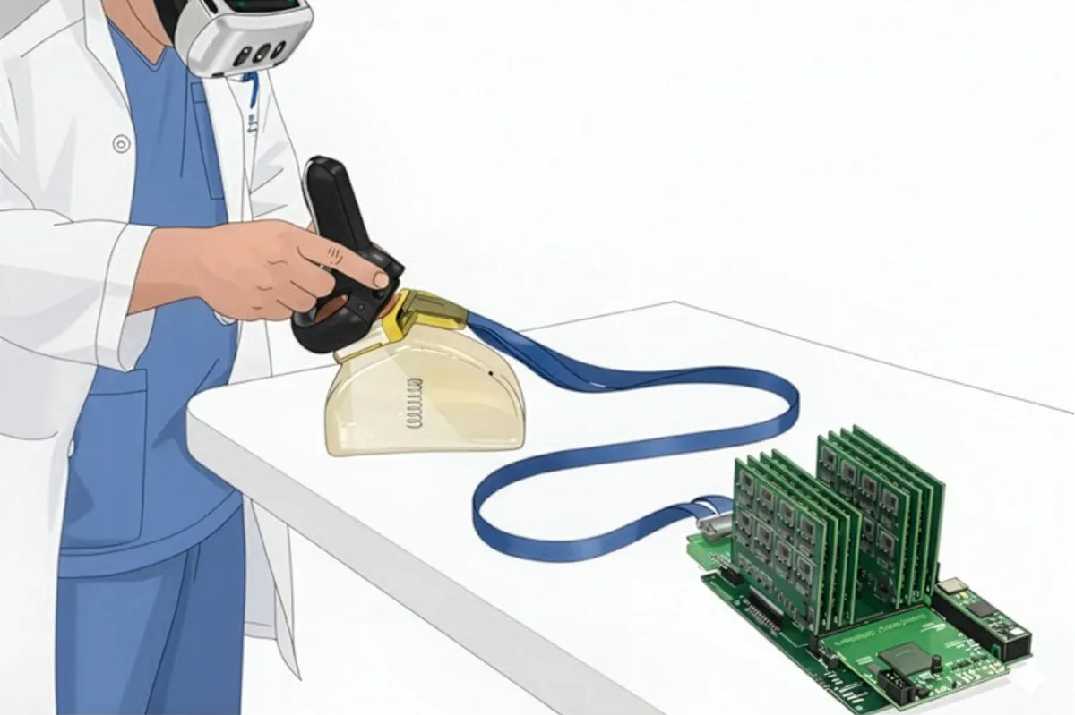
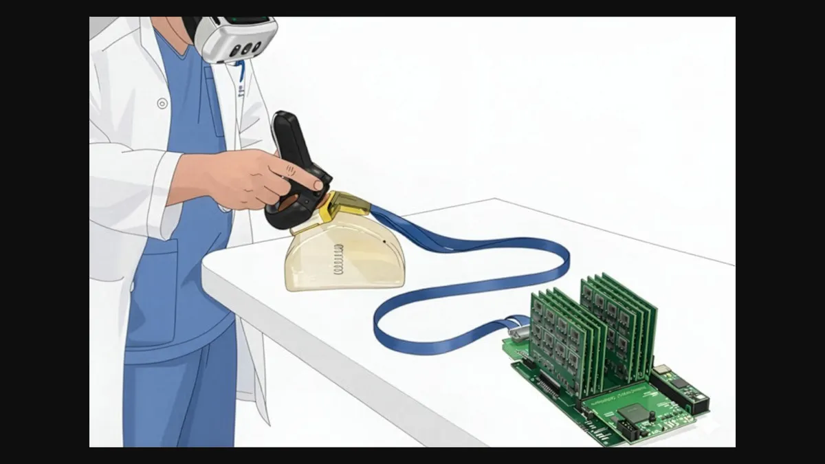
The MIT team combined a real-time 3D ultrasound system (one they initially developed for breast cancer detection) with augmented reality. They built a specialized, compact ultrasound probe with an empty square array, allowing it to capture 3D images. Crucially, this system uses fewer ultrasound elements than typical 3D setups, making it cheaper and less power-hungry. Because even X-ray vision needs to be practical.
All that juicy 3D ultrasound data is then streamed into a 3D computer graphics engine (yes, the same kind that powers video games like Fortnite). This engine converts the data into a direct 3D representation, which then appears superimposed over the actual object when viewed through an AR/VR headset. You can tilt your head, move around, and essentially peer into the body as if it were transparent.
They tested AR-VIU with 18 participants: nine ultrasound experts and nine complete novices. The task? Identify objects (like a screw in gelatin) or mark a "tissue phantom" (a material mimicking human tissue) for a simulated biopsy.
The results were pretty definitive: AR-VIU vastly improved everyone's ability to identify and locate objects. Novices, using AR-VIU, performed almost as well as experts using the traditional 2D method. Let that sink in for a moment. Novices. Almost as good as seasoned pros. Because apparently, seeing in 3D is easier than guessing in 2D.
While some experts, set in their ways, still preferred their old 2D screens, they readily acknowledged AR-VIU's potential for high-stakes tasks like needle placement or visualizing heart movement. Because even the best of us can appreciate a little help from technology, especially when it involves not having to mentally reconstruct a human organ. This isn't just about making things easier; it's about making them clearer, faster, and, dare we say, a bit more sci-fi.