
Turns out, the secret to your crystal-clear central vision isn't just about what your eyes grow, but what they change into. Scientists at Johns Hopkins University just flipped an old theory on its head, discovering a precise prenatal dance between vitamin A and thyroid hormones that sculpts our sharpest sight.
This isn't just a fun fact for your next dinner party. It’s a discovery that could pave the way for entirely new treatments for vision loss from villains like macular degeneration and glaucoma. Because apparently, even something as fundamental as how we see can still hold decades-old surprises.
The Tiny Spot That Sees It All
Robert J. Johnston Jr., a biology professor at Johns Hopkins, led the charge. He explains that understanding the very center of the retina—the foveola—is critical. It's the MVP of your vision, handling about half of everything you see, and it’s often the first casualty in diseases like macular degeneration. Get this area right, and you might just grow and transplant new tissue to restore sight.
We're a new kind of news feed.
Regular news is designed to drain you. We're a non-profit built to restore you. Every story we publish is scored for impact, progress, and hope.
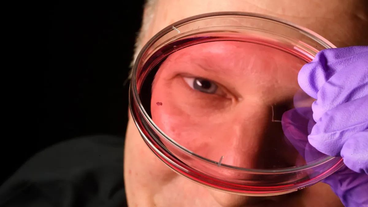
Start Your News DetoxTo crack the code, researchers didn't just stare at eyeballs. They grew them. Or rather, they grew retinal organoids: tiny clusters of tissue from fetal cells that mimic parts of the retina. They watched these lab-grown eyes for months, charting the cellular drama that eventually forms the foveola. This minuscule region gives us our sharpest, most detailed vision.
The foveola's secret? It's packed only with red and green cone photoreceptors. These are the light-sensing cells that give us daytime and color vision. Humans are unique with three types of cones (blue, green, red), allowing for a dazzling spectrum of color perception. But how the foveola ends up with just two of those colors has been a decades-long head-scratcher. Mice and fish, bless their hearts, just don't have the same setup.
The Great Cone Conversion
The new findings reveal a prenatal plot twist. Around weeks 10 to 12 of fetal development, a few blue cones pop up in the foveola. But by week 14, plot twist number two: these blue cones transform into red and green cones. Talk about a career change.
This cellular metamorphosis happens in two acts. First, retinoic acid (a derivative of vitamin A) breaks down, reducing the production of new blue cones. Then, thyroid hormones swoop in, prompting the existing blue cones to switch teams and become red or green cones. Johnston explains that retinoic acid sets the stage, and thyroid hormone performs the conversion. Why? Because blue cones in that tiny, critical area would actually reduce vision quality. Who knew color perception was so picky?
This discovery directly challenges a theory that's been gospel for about 30 years. The old idea was that blue cones formed in the center and then simply moved out of the way. Like they decided, "Nah, this isn't my scene," and relocated. But the new evidence suggests they just stay put and change their identity entirely. Which, if you think about it, is both impressive and slightly terrifying. Imagine if your phone could just decide to become a toaster.
A Clearer Future?
This isn't just academic musing. These insights could lead to entirely new strategies for treating vision loss. Johnston’s team is now busy refining their retinal organoids, hoping to make them even more human-like. Better models mean better understanding, which means healthier photoreceptor cells for future cell replacement therapies. We're talking about a potential cure for macular degeneration, a disease that currently has no good options.
Katarzyna Hussey, a molecular and cell biologist at CiRC Biosciences, notes that the ultimate goal is to create custom photoreceptor populations using this organoid tech. The long game? Introducing healthy cells that can integrate into the eye and restore lost vision. It's a long road of safety and effectiveness studies, but it’s a path that suddenly looks a whole lot clearer.