A Zoom call during lockdown led to a breakthrough that's reshaping how we see the microscopic life sustaining our oceans. For decades, scientists could read the genetic code of plankton but couldn't actually see inside them — their cell walls were impenetrable barriers. Now, a technique called expansion microscopy is changing that.
Plankton are the foundation of ocean life. These single-celled organisms produce roughly half the oxygen we breathe and feed everything from fish to whales. Yet we've known surprisingly little about their internal structure. The problem wasn't lack of interest. It was a practical one: the microscopes powerful enough to see inside these cells didn't exist, and the cells themselves refused to cooperate with the techniques scientists tried.
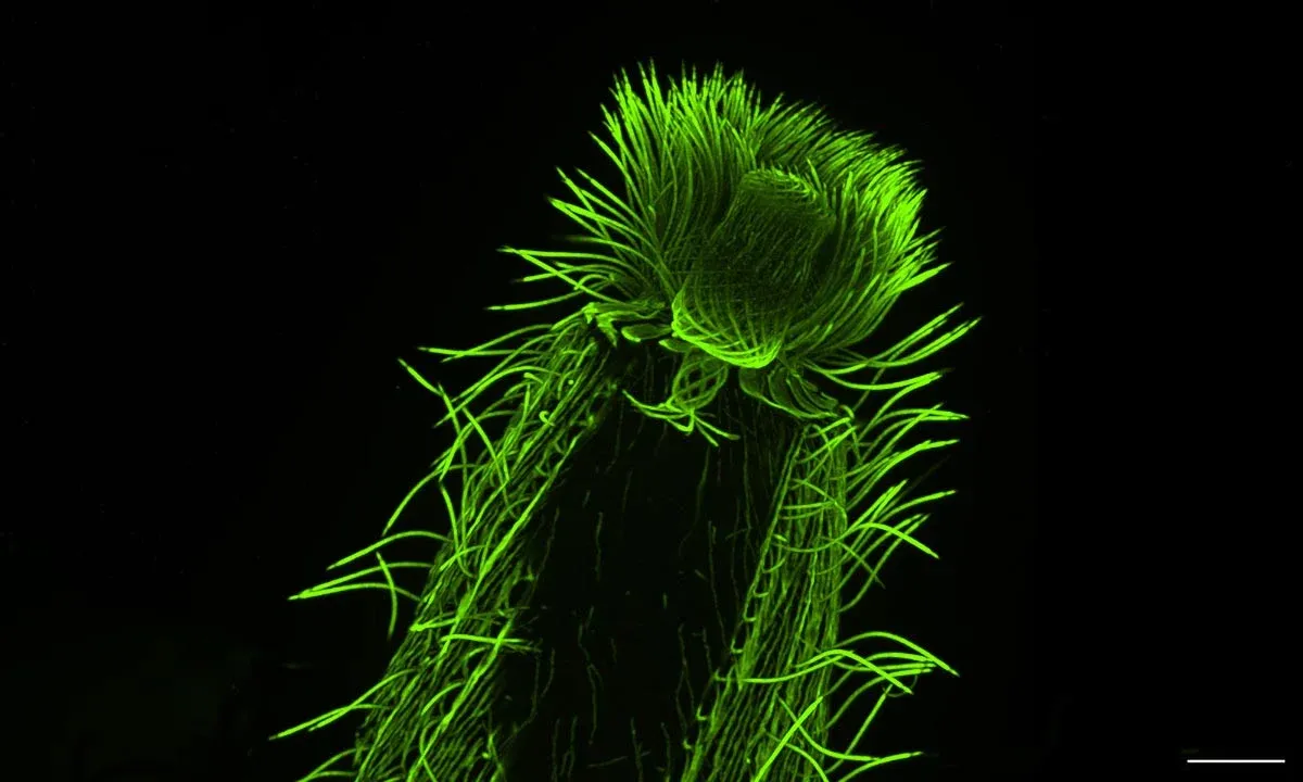
When EMBL researcher Gautam Dey got that pandemic-era call from collaborator Omaya Dudin at the Swiss Federal Institute of Technology, Dudin had news. He'd refined a technique that could finally crack the problem. Expansion microscopy works by embedding a sample in a gel that absorbs water and expands — stretching the cell up to 16 times larger while keeping its internal structures intact. It's like inflating a tiny balloon without popping it.
We're a new kind of news feed.
Regular news is designed to drain you. We're a non-profit built to restore you. Every story we publish is scored for impact, progress, and hope.
Start Your News DetoxSeeing What Was Always Hidden
The technique itself wasn't brand new. MIT scientists had developed it a decade earlier, and researchers at the University of Geneva had adapted it specifically for looking at cellular ultrastructure. But applying it to plankton — creatures with notoriously stubborn cell walls — was the breakthrough.
Dey, Dudin, and colleagues Paul Guichard and Virginie Hamel decided to push harder. They partnered with a European research expedition called TREC (Traversing European Coastlines) that was already collecting marine samples. Their first major sampling site was Roscoff, France, home to one of Europe's largest collections of marine microorganisms. When they asked the facility's manager how many samples might be available for testing, they expected around 20. Instead, they got access to more than 200.
"We spent three days and nights just fixing those samples," recalled Felix Mikus, one of the researchers. "This was a treasure trove we could not let go of."
That work paid off. In a study published in Cell this year, the team published detailed views of the internal architecture of over 200 plankton species. They focused especially on the cytoskeleton — the filament network that gives cells their shape — mapping how microtubules and related proteins are organized across different species.
What they found matters for understanding evolution. By comparing the internal structures across many species, they could trace how cellular architecture has changed over millions of years. Dinoflagellates, one of the ocean's most diverse groups, showed particular variation in how they organize these structures — clues about how they adapted to different ocean environments.
The Next Map: A Planetary Atlas
The team has already secured funding to keep going. A $2 million grant from the Moore Foundation will support the next phase: building what Dey calls a "planetary atlas of plankton." The goal is ambitious — to map the internal structure of hundreds or thousands of plankton species and link that cellular architecture to genetic data, creating a comprehensive picture of how microscopic ocean life is built.
This matters because plankton aren't just background players. They're the foundation of ocean food webs, carbon cycles, and oxygen production. Understanding their structure — how they're organized, how they move, how they divide — is understanding how life itself works at the smallest scale. And it's understanding the ocean's most fundamental workers in a way that was impossible just a few years ago.