New research from Johns Hopkins University is changing how we understand eye development. Scientists have found that sharp, color vision begins before birth. This happens through a team effort between a molecule from vitamin A and thyroid hormones in the retina.
This discovery challenges ideas that have been around for a long time about how the eye's light-detecting cells form. It could also help future research into treatments for vision problems like macular degeneration and glaucoma.
How Vision Develops
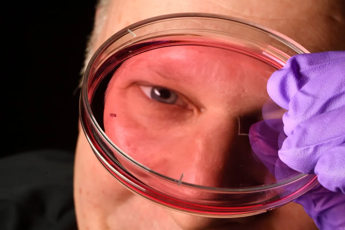
The study used lab-grown retinal tissue, called organoids. These are small groups of tissue grown from fetal cells. By watching these lab-grown retinas for several months, researchers saw how the foveola forms. The foveola is a key part of the retina that gives us sharp vision.
We're a new kind of news feed.
Regular news is designed to drain you. We're a non-profit built to restore you. Every story we publish is scored for impact, progress, and hope.
Start Your News DetoxRobert J. Johnston Jr., a biology professor at Johns Hopkins, led the research. He explained that understanding this part of the retina is crucial because it's the first area to fail in people with macular degeneration. The hope is to one day grow and transplant these tissues to bring back vision.
The study looked at photoreceptors, which are cells that help us see in daylight. These cells become blue, green, or red cone cells, each sensitive to different light colors.
The foveola is tiny, but it handles about 50% of what we see. It has red and green cones, but no blue cones. Blue cones are spread out more across the rest of the retina. Humans are unique because they have three types of cones, allowing for a wide range of color vision.
A New Understanding of Cone Cells
Researchers found that the way cones are spread in the foveola comes from a series of events early in development.
Around weeks 10 to 12, a few blue cones appear in this area. But by week 14, these blue cones change into red and green cones. The study points to two main reasons for this change. First, a molecule from vitamin A, called retinoic acid, breaks down. This limits how many blue cones form. Second, thyroid hormones help turn the existing blue cones into red and green ones.
Johnston noted that retinoic acid helps set the pattern, and then thyroid hormone converts the remaining cells. This is important because having blue cones in that area would make vision less clear.
Challenging Old Theories
These findings go against a 30-year-old idea that blue cones simply move away from the foveola as the eye develops. Instead, the new research suggests these cells actually change their identity to get the right mix of cone types.
Johnston explained that the old model suggested cells decide what they will be and stay that way forever, or that blue cones just move out. He said their data supports a different model: these cells convert over time, which is very surprising.
This new understanding could lead to new ways to treat vision loss. Johnston's team is working to make their organoid models even better, so they act more like a real human retina.
Katarzyna Hussey, a study author and former doctoral student, said this progress could help create better photoreceptors. Eventually, it might lead to cell-based treatments for diseases like macular degeneration, which currently have no cure.
Hussey, now a molecular and cell biologist, believes that using organoid technology could eventually allow them to create custom populations of photoreceptors. This could lead to cell replacement therapy, where healthy cells are introduced to the eye to restore lost vision. She added that while these are long-term goals, it's a promising path forward.
Deep Dive & References
A cell fate specification and transition mechanism for human foveolar cone subtype patterning - Proceedings of the National Academy of Sciences, 2026