MIT researchers have made a breakthrough in laser technology. They found that a chaotic laser beam can unexpectedly organize itself into a precise "pencil beam." This discovery allows scientists to watch drugs enter the brain in real time, much faster and with high detail.
This new method could help test treatments for diseases like Alzheimer's or ALS. It shows if drugs successfully cross the blood-brain barrier and reach their targets.
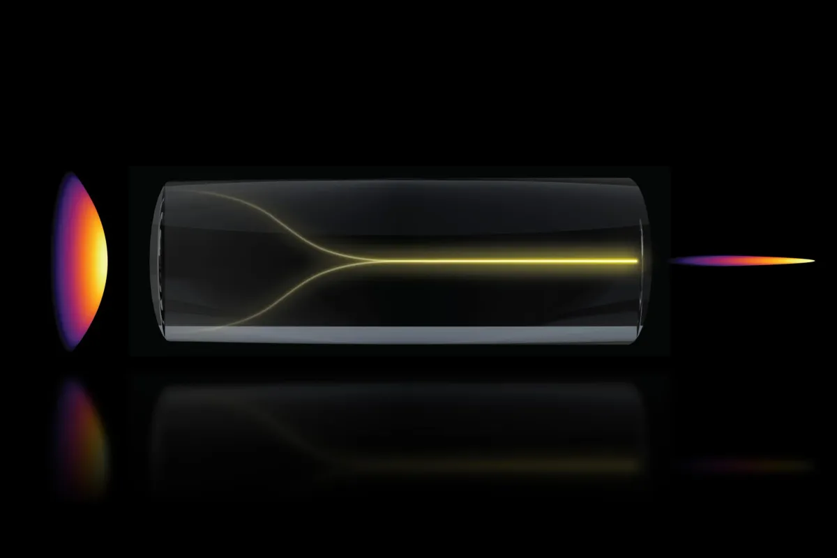
How the Laser Beam Self-Organizes
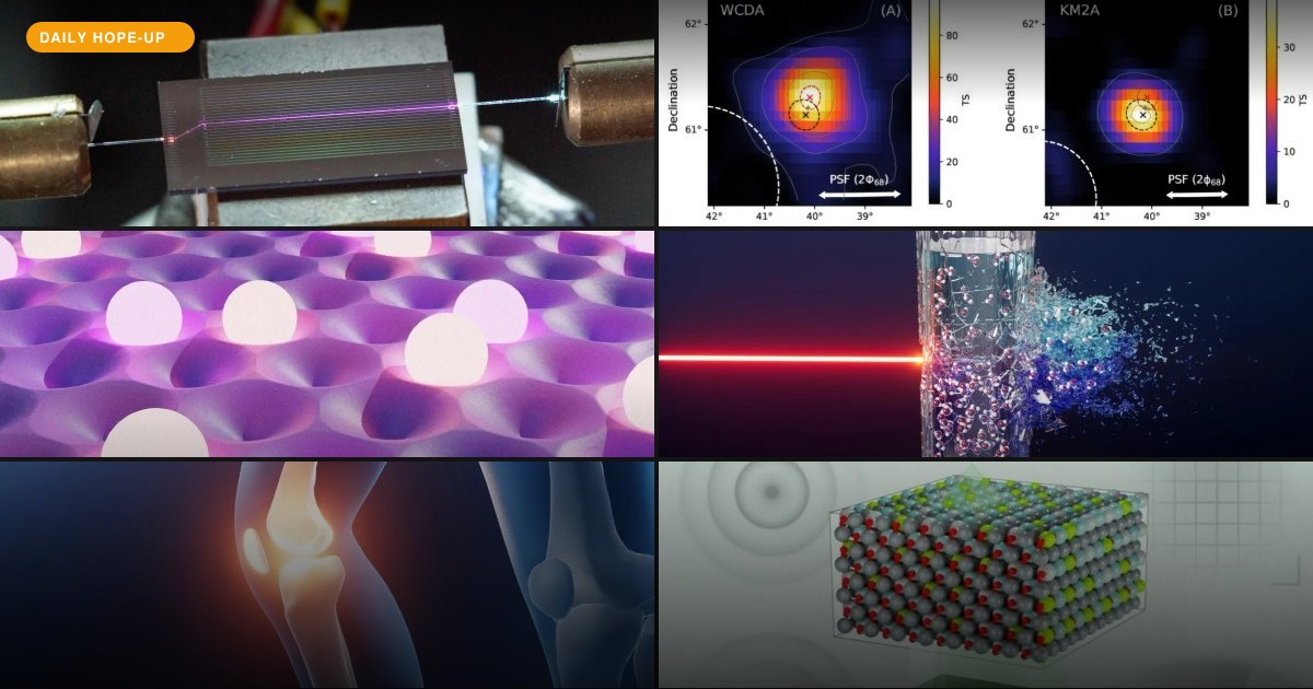
The discovery started with a puzzling observation. Researchers were testing how much power a special optical fiber could handle. They expected the laser light to become more scattered and disordered at higher power. Instead, as the power got very high, the light suddenly focused into a single, sharp beam.
We're a new kind of news feed.
Regular news is designed to drain you. We're a non-profit built to restore you. Every story we publish is scored for impact, progress, and hope.
Start Your News DetoxSixian You, an MIT assistant professor, explained that the common belief was that high power would make the light chaotic. But their work showed that light can organize itself into a new solution for bioimaging.
To make this happen, two things are needed. First, the laser must enter the fiber perfectly straight, at a zero-degree angle. Second, the power must be high enough for the light to interact directly with the fiber's glass.
Honghao Cao, a lead author on the study, noted that at this critical power, the light's "nonlinearity" balances the fiber's natural disorder. This creates a stable pencil beam without needing special equipment.
These conditions are not usually explored because researchers avoid high power to prevent fiber damage. Also, such precise alignment is not typically considered necessary for multimode fibers.
Faster, Clearer Brain Imaging
The new pencil beam is stable and provides high resolution. Unlike many traditional beams, it doesn't create blurry halos that can reduce image clarity. This makes it ideal for biomedical imaging.
The team used the technique to image the human blood-brain barrier. This barrier protects the brain but also blocks many medications. Scientists need to track how drugs cross it.
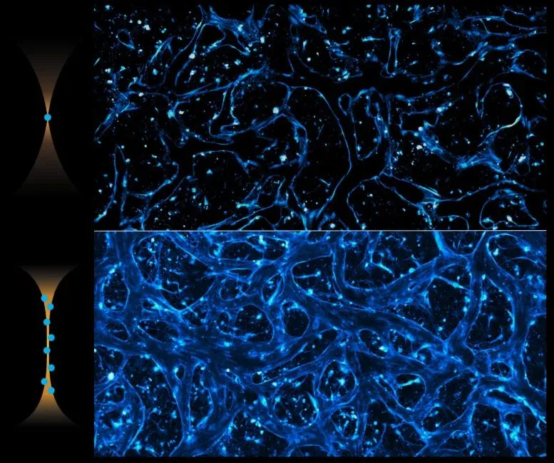
Traditional methods capture one 2D image slice at a time, then combine them. The new pencil beam method creates 3D images about 25 times faster, with similar quality. It also lets scientists watch cells absorb proteins in real time.
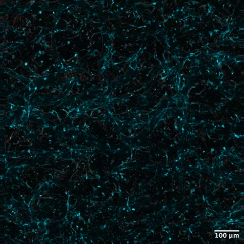
Roger Kamm, an MIT professor, highlighted that this method doesn't require fluorescent tags on cells. This is a big step forward for the pharmaceutical industry. It allows them to see how drugs enter the brain and how quickly different cell types take them in.
This approach can also be used for other engineered tissue models. It offers a powerful tool for biological engineering by tracking various compounds and molecular targets over time.
The researchers achieved cellular-level 3D images with better quality and much faster speeds. You explained that this method overcomes the usual trade-off between image resolution and how deep you can see.
What's Next
The team plans to study the physics behind this self-organizing beam more deeply. They also want to use it for other applications, like imaging neurons in the brain, and explore ways to bring it to market.
Deep Dive & References
- Self-localized ultrafast pencil beam for volumetric multiphoton imaging - Nature Methods, 2026