
Researchers have created a new high-speed microscope. It can image large, moving samples in great detail.
For a long time, microscope designers had to choose between speed, how much area they could see, and how clear the image was. Improving one usually meant making another worse. Now, a team led by UC Berkeley has found a way around this problem. This opens up new possibilities for microscopy.
A Breakthrough in Computational Microscopy
The researchers built a computational microscope that can see details as small as microns across areas several centimeters wide. It does this faster than video speed. The new microscope uses 48 camera sensors and special computational imaging methods. It can capture videos at 25.2 billion pixels per second. This is a new record for combining high detail, a wide view, and fast imaging.
We're a new kind of news feed.
Regular news is designed to drain you. We're a non-profit built to restore you. Every story we publish is scored for impact, progress, and hope.
Start Your News DetoxLaura Waller, a professor of electrical engineering and computer sciences and the study's lead investigator, called this a "breakthrough." She explained that their microscope achieved 3-micron resolution over 5 square centimeters at 120 frames per second. This is not possible with traditional microscopes.
Waller noted that their method could image many live organisms at once and watch samples over time.
Overcoming Imaging Limits
Optical engineers have struggled with limits on how much spatial and temporal information a microscope can capture at the same time. This means it's hard to get both fine details (high resolution or a wide view) and fast movement (high frame rate). These limits make it difficult to study dynamic samples, like organisms moving freely.
To solve this, the researchers combined a camera sensor array, a special optical element, and an optimization algorithm. This allowed them to capture gigapixel-scale images instantly.
Their new microscope uses 48 separate sensors on a circuit board about the size of a credit card. When in use, these sensors act like one giant sensor. This increases the number of pixels captured and improves image quality.
However, there's a challenge: gaps between the sensors mean some light and data are missed. This makes it hard to reconstruct a complete image.
To fill these gaps, Waller explained they used "compressed sensing tricks." They made a custom glass plate that redirects light that would normally fall between the sensors onto them instead. Then, a computational algorithm fills in the missing data and reconstructs the image.
Imaging Static and Dynamic Samples
The researchers tested their microscope with both static and moving samples. For static samples, their reconstructed images matched low-resolution traditional images in overall features but showed much higher detail.
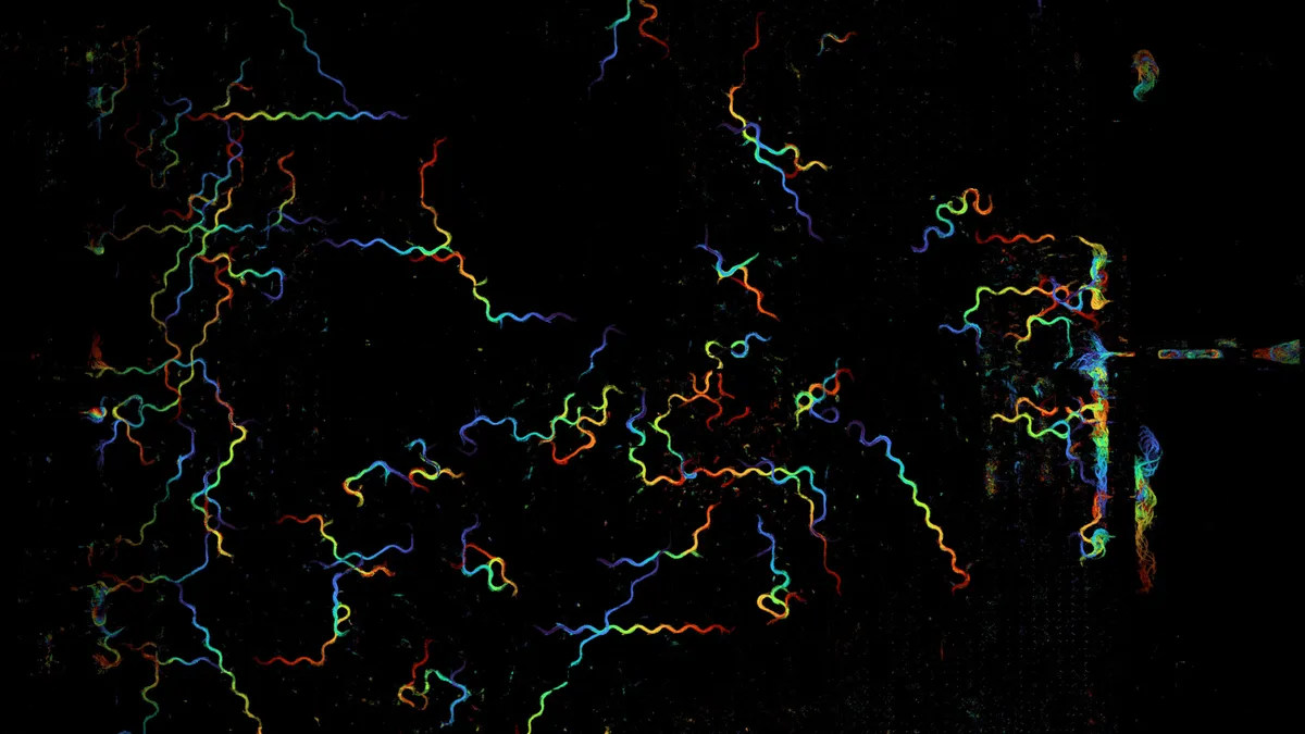
For dynamic samples, they imaged dozens of C. elegans nematodes moving freely. They captured these worms at 120 frames per second for 15 seconds.
Kevin C. Zhou, the lead author, said the microscope's ability to combine high resolution, a wide view, and high speed allowed them to track the C. elegans worms and even structures inside them. For example, they could track individual worms and observe their rapid pharyngeal pumping, which is part of their feeding behavior.
The researchers also noted that their computational method removed the need for the difficult manual calibration usually required for such a large microscope. This saves a lot of time and effort. Co-author Chaoying Gu believes that "calibration-free capabilities like our method will prove to be one of the key ingredients for scaling up imaging systems in the future."
Waller thinks this work will help advance computational microscopy. She stated that "computational imaging — the joint design of hardware and software — has a lot to offer in scaling up to large data acquisition."
Deep Dive & References
Computational gigapixel microscopy for high-throughput cellular imaging - Nature Photonics, 2026