
For years, scientists have been staring at most human proteins and basically shrugging. They were just too darn small for even the most powerful microscopes to properly see. Imagine trying to identify a specific grain of sand from orbit. That was the situation.
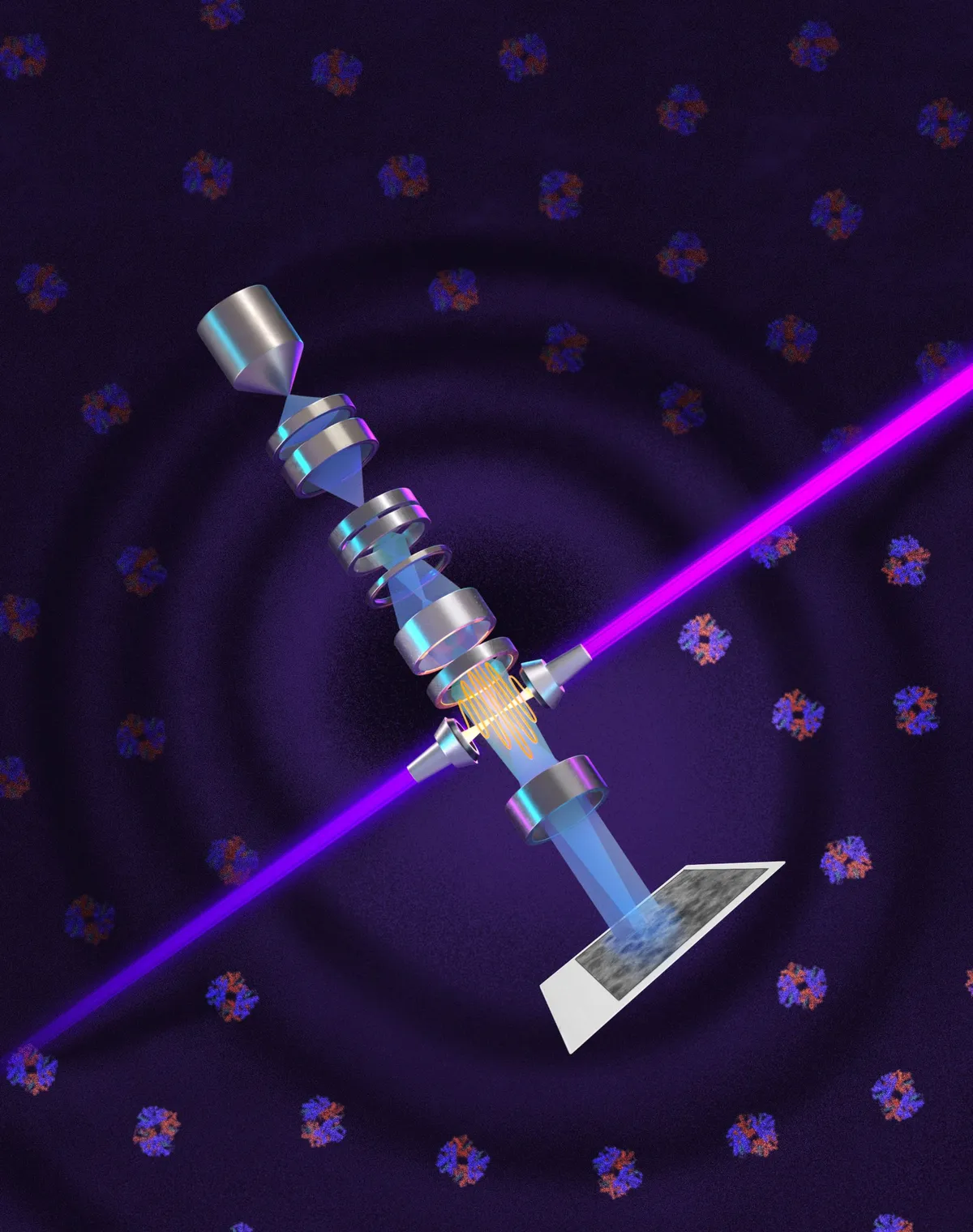
Now, physicists at UC Berkeley have cooked up a laser-based system that’s about to change all that. They’ve tweaked an old imaging trick called phase contrast and bolted it onto cryoelectron microscopes (cryo-EM), creating what they've dubbed a "laser phase plate." The result? Clearer images, without weakening the electron beam that does all the heavy lifting.
This isn't just about pretty pictures. It could supercharge cryoelectron tomography (cryo-ET), which stitches together images from different angles to build 3D models of molecules inside cells. Because apparently that’s where we are now. We're building tiny 3D maps of the stuff that makes us, us.
We're a new kind of news feed.
Regular news is designed to drain you. We're a non-profit built to restore you. Every story we publish is scored for impact, progress, and hope.
Start Your News DetoxTheia, Goddess of Light (and Tiny Proteins)
Holger Müller, a UC Berkeley physics professor, led this microscopic revolution. He explains that cryo-EM is the go-to for mapping biological molecules, while cryo-ET wants to see how those molecules dance together inside a cell. The snag? Many human proteins are just too petite for current tech to handle. The laser phase plate, Müller hopes, will boost the signal-to-noise ratio, making those shy proteins pop.
His team built a special instrument they named Theia, after the Greek goddess of light. It's essentially a souped-up Thermo Fisher Krios microscope with the laser phase plate added. Müller calls it a "Formula 1 microscope," even without the laser. Add the laser, and he expects it to become the best instrument in the world. Which, if you think about it, is both impressive and slightly terrifying.
Biohub, who funded Theia, is already eyeing a second microscope with two lower-power lasers, aiming to reduce distortions and protect the delicate parts. Because even Formula 1 microscopes need a pit crew.
The Nobel-Winning Idea, Reimagined
The core idea behind phase contrast isn't new; it dates back to Dutch physicist Frits Zernike in 1930. He realized that light passing through biological samples changed in both brightness and phase. By subtly shifting the unscattered light by 90 degrees, he could turn those tiny, invisible phase differences into clear, visible brightness changes. No stains needed. Zernike snagged a Nobel Prize in 1953 for that one.
Scientists tried to adapt it for electron microscopes, but early attempts were… messy. They weakened the beam, blurred images, or just made everything unstable. Then, in 2010, Müller and cryo-EM guru Robert Glaeser suggested a powerful laser could be the answer.
Fifteen years later, Müller’s team has made it work. They trapped a laser inside a mirrored cavity, where the light bounces over 10,000 times, focusing into a spot more intense than what you’d use for welding. Because when you’re trying to see the invisible, you don’t mess around.
Tests on six biological samples showed the laser really shone with the smallest, most challenging specimens. Take hemoglobin, for instance — a protein so small it usually hides from current instruments. The laser phase plate made it significantly clearer. Suddenly, we're seeing things that were once just theoretical blurs.
What This Means for You (and Your Proteins)
Currently, cryo-EM struggles with proteins smaller than about 70 kilodaltons. The problem? About 90% of human proteins are smaller than that. We’ve been missing most of the party.
With the laser phase plate, researchers can now image proteins as small as 50 kilodaltons, which is a massive leap. Müller hopes future tweaks will push that limit down to 17 kilodaltons, opening up a whole new universe of biological understanding.
Stephani Otte, Biohub’s VP of Imaging Science, calls this a "major change for biology." It means scientists could finally see how the molecular machinery inside our living cells actually works, not just what it looks like. And that, in turn, could fundamentally shift how we understand and tackle diseases. Because if you can finally see the problem, you might just be able to fix it.