
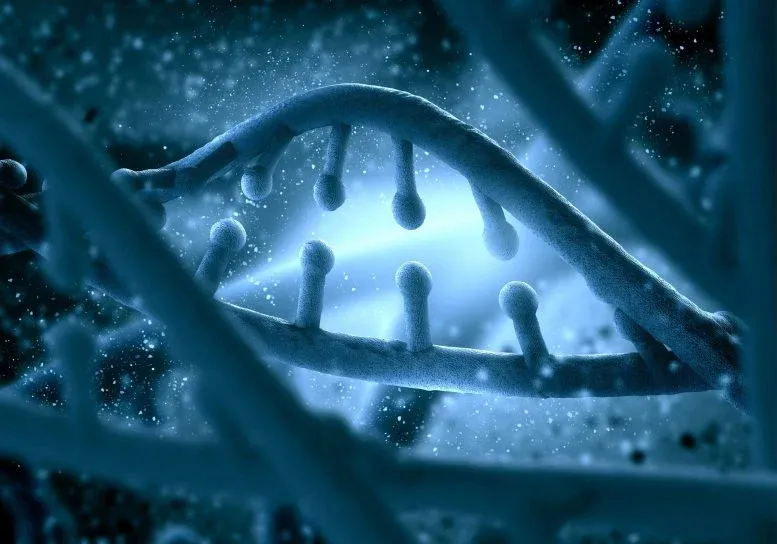
Imagine trying to untangle two meters of spaghetti and then stuffing it all into a thimble. That's essentially what a cell does with your DNA. Now, scientists have developed new tech that lets them see this microscopic origami happening in real-time, at a resolution so sharp it's practically peeking at the individual DNA strands.
This isn't just a cool party trick for microscopes. It's a breakthrough that could fundamentally change how we diagnose and treat diseases, especially cancer. Turns out, how your DNA is packed might be a huge clue to what's going wrong inside your body.
The Blinking Probes That Changed Everything
For years, getting a clear picture of DNA folding in living cells was like trying to photograph a ghost in a hurricane. Powerful microscopes needed harsh chemicals and intense lasers, which, predictably, aren't great for living cells. Most images came from dead cells, which, while informative, don't show the dynamic dance of DNA in action.
We're a new kind of news feed.
Regular news is designed to drain you. We're a non-profit built to restore you. Every story we publish is scored for impact, progress, and hope.
Start Your News DetoxEnter the new fluorescent probes, cleverly named HoTs. These tiny dyes can slip into living cells, bind to DNA, and then — here's the magic — blink on and off. Why blink? If all the dyes glowed constantly, their signals would just blur into a mess. But by detecting individual probes as they momentarily switch on, an advanced microscope can pinpoint their exact location. A computer then stitches together thousands of these fleeting snapshots to create an image ten times sharper than a regular microscope. Pretty clever for something the width of a DNA double helix.
They tested these HoTs on preserved bowel tissue from cancer patients. The results were stark: DNA in tumors looked looser, more spread out, compared to healthy tissue. Other studies have hinted at this, suggesting that DNA unpacks as cancer progresses. This new, super-resolution view could offer a crucial clue to a tumor's aggressiveness.
AI Gets in on the Action
Not content with just seeing, the researchers also brought in AI. They used AINU, a program developed in 2024, which is designed to spot subtle patterns in nuclear DNA that are invisible to the human eye. After training AINU on these new, super-sharp images of living cells, the program could distinguish skin cells from stem cells with 96% to 98% accuracy. Think about that: two cell types with identical DNA, but the AI could tell them apart just by how their DNA was folded.
This is huge, because AINU previously only worked with dead cells. Now, it can analyze living ones, potentially helping doctors differentiate cancerous tissue from healthy tissue, or even identify the most promising stem cells for regenerative medicine. Which, if you think about it, is both impressive and slightly terrifying.
While the sharpest images are still coming from preserved cells, the team is already working on getting the same precision in living ones. Because apparently, seeing DNA in near-molecular detail is just the beginning. Your dinner table stories just got a lot more microscopic.